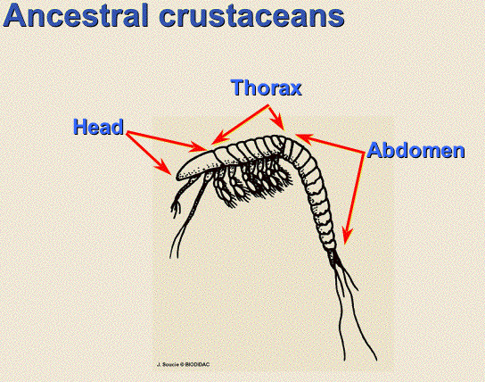
Ancestrally arthropods had two tagmata; the head and trunk with a pair of legs on each of the trunk segments.
Appendages on the head include antennae and feeding appendages; chelicerae and mandibles for example. On the trunk, each leg was involved in locomotion, gathering food and gas exchange and you can still see this original organization in Crustacea. But the secret of the group is how appendages on different segments became specialized for different functions, or disappeared completely. For example appendages that became specialized for only locomotion required larger muscles and the shape and size of the segments with walking leg changed to accommodate the musculature and how the plates of cuticle moved relative to each other. Adjacent segments with walking legs became a tagma, in this case a thorax, and the process is referred to as tagmosis. As we look at the groups. you should be keeping track of the different tagmata and the appendages that are attached to them.
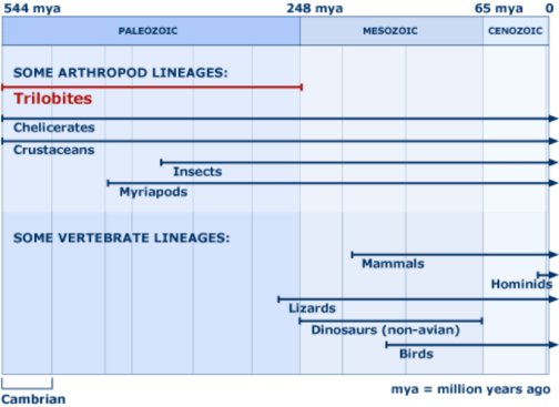
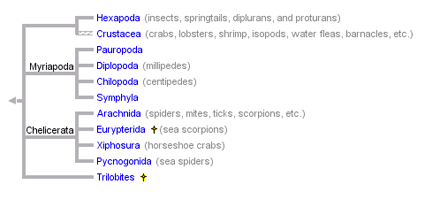
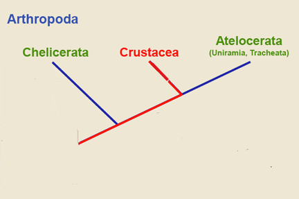
The current phylogeny for arthropods
This is a simpler type of scheme than presented by your text. There is a move to make this scheme even simpler and resolve the problem with the clade Crustacea by collapsing all arthropoda into three big clades.
Classes (Evolutionary scheme) or major clades:
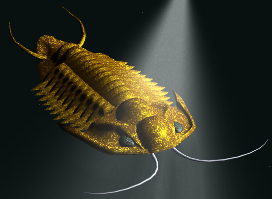
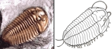
1. Trilobita or simply trilobites: extinct, An early branch of arthropod life, the trilobites, underwent great diversity in the period roughly spanning 400-200 million years ago. They dominated the aquatic worlds of that time much as insects dominate terrestrial life today.
Trilobites are the most diverse group of extinct animals preserved in the fossil record. Exoskeletons were highly calcified, and thus well preserved
Trilobites were extremely successful, found in a very wide variety of ocean habitats, and probably occupied many, if not all of the ecological niches that crustaceans do today.http://www.trilobites.info/feeding.htm
bottom dweller
pelagic
When extinct about 251 million years ago (Permian) when > 90% of all species on the planet were extinguished.
Trilobites were a very successful group and a diverse group even in the Cambrian. So how long did trilobites exist? Longer than the dinosaurs.
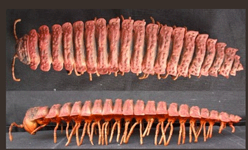
Body divided into three lobes, two different ways: Antero-posteriorly into a cephalon (head, containing most of the viscera), thorax (generally with many segments, containing the limbs), and pygidium (fused "tail" segments)
Medio-laterally into a central axial lobe (containing the nervous and digestive systems) and a pair of lateral pleural lobes (overlying the spread of the limbs, including their gills).
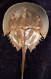
2. Merostomata (classic) or Xiphosura (cladistic), or horseshoe crabs (common)
Body divided into two distinct portions with no distinct head. No antennae and first pair of appendages are adapted for feeding. Makes it part of the clade Chelicerata
Distinguished by book gills and telson (class distinctions).
Aquatic (most marine, but a few fossil brackish and freshwater forms), but can come onto land for short periods of time.
Fossil record from Silurian (445 million years ago) onward
Early forms and larvae of modern forms are VERY trilobite-like.
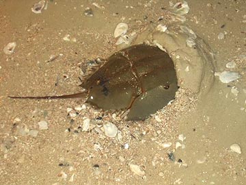
Horseshoe crabs feed on the sea bottom, capturing small molluscs and worms with their pincers. They break up hard material, such as mollusc shells, by grinding it between the hard edges of their upper legs or the gnathobases. The chelicerae, or the chilaria, small degenerate legs located behind the pusher legs, can push any food into the crab's mouth. A horseshoe crab also has a gizzard that contains sand and small bits of gravel to help grind its food.
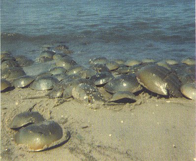
In horseshoe crabs, fertilization is external. During the spring breeding season, in the North American species, thousands of the animals come ashore to breed in shallow water. Each male attaches to a female and is dragged around until she is ready to lay eggs. Young horseshoe crabs return to the water as free-swimming larvae.
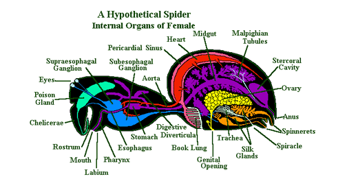
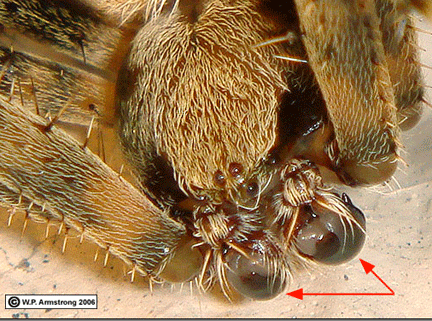
Two main body parts: the prosoma (also called the cephalothorax) and the abdomen (also called the opisthoma).The first two appendages are the chelicerae and pedipalps which are both involved in feeding. The chelicerae tear apart food. Next pair is the pedipalps which are modified for grabbing, killing or reproducing. Immediately behind these are the characteristic four pairs of walking legs. The second tagma, opisthosoma, has no distinct appendages, although the spinnerets used to make silk are probably the remnants of the ancestral appendages on the opisthosoma.
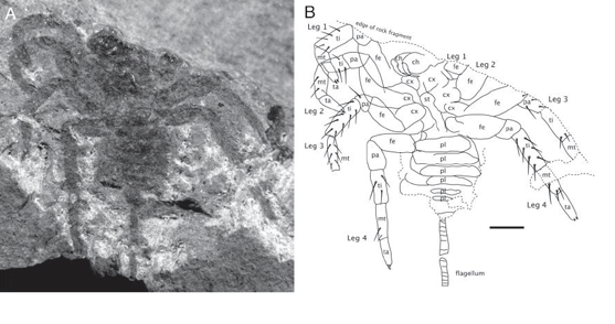
The oldest known arachnid is the Palaeotarbus jerami, from about 420 million years ago in the Silurian period, and had a triangular Cephalothorax and segmented abdomen, as well as eight legs and a pair of pedipalps. Attercopus fimbriunguis from 386 million years ago in the Devonian period, bears the earliest known silk-producing spigots, and was therefore hailed as a spider. However these spigots may have been mounted on the underside of the abdomen rather than on spinnerets, which are modified appendages and whose mobility is important in the building of webs. Hence Attercopus and the similar archnid Permarachne may not have been true spiders, and probably used silk for lining nests or producing egg-cases rather than for building webs.
Attercopus fossils
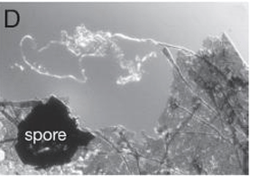
Due to the fact that spiders' bodies are quite soft, the vast majority of fossil spiders have been found preserved in amber. The oldest known amber that contains fossil arthropods dates from130 million years ago in the Early Cretaceous period. In addition to preserving spiders' anatomy in very fine detail, pieces of amber show spiders mating, killing prey, producing silk and possibly caring for their young. In a few cases amber has preserved spiders' egg sacs and webs, occasionally with prey attached.
True Spiders (old order Araneae)
Most spiders breathe through organs known as book lungs. Book lungs consist of an atrium or space into which numerous layers of membrane bound tissue extend. Hemolymph flows through these thin layers of tissue and gaseous exchange occurs across the membrane. The overall effect is of a series of sheets of tissue with air spaces between each sheet. This arrangement means that there is a large surface area here, up to 70 sq. cms. in large tarantulas, across which gaseous exchange can occur.
In the primitive spiders, there are two pairs of book lungs, however in more modern spiders one of these pairs has become modified into a pair of tubular tracheae which branch out throughout the whole body. In a few families the second pair of book lungs has also been modified or lost. Scientists agree that the book lung system is older system and that tracheae developed later.
Spiders may use malphigian tubules or coxal glands for excretion.
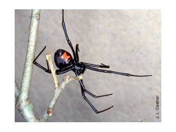
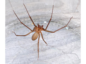
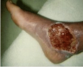
Spiders are notorious for their venom. The Black Widow Spider is responsible for occasional deaths, and the bite of a Brown Recluse can cause an ulcerating wound that refuses to heal. Most spiders however, are harmless to humans.
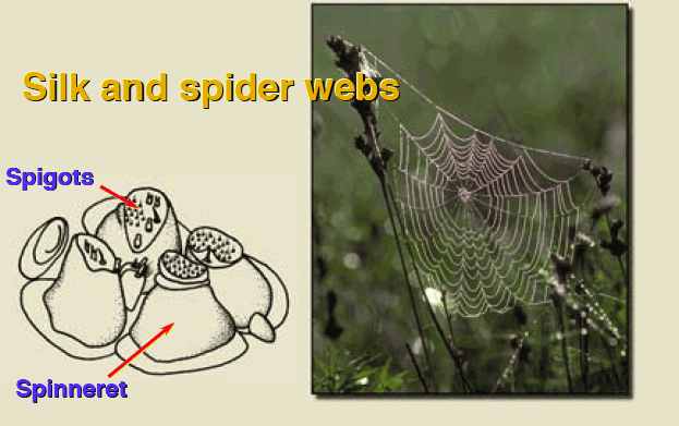
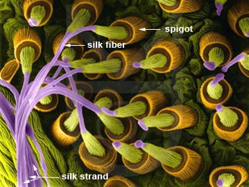
Spiders can produce silk by secretions of an abdominal gland, and many spiders use this sticky material to construct a web.
The glands on the Madagascar spider's (Nephila madagascariensis) right side. There are glands on the left side as well. Silk glands 1 and 2 produce the dry silk the spider holds on to when walking on its web, or when climbing up and down. The viscid silk is produced in another gland (3). This basic silk is coated by the adhesive (sticky) glands (4 and 5). The 6th gland produces the adhesive necessary for sticking the silk to another surface. The 7th gland produces the raw material for a special thin silk used to wrap the prey up after it is caught. The 8th gland produces the silk for the cocoon. 9, 10, and 11 show the back, central, and front spinnerets (silk nozzles).
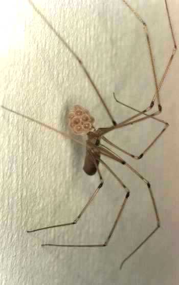
Spiders perform an elaborate courtship ritual. In web spiders, the male is much smaller than the female and must approach cautiously to avoid becoming prey. If successful, he mounts the female and uses his pedipalps to deposit sperm into her sperm receptacles. Most female spiders guard their eggs and some in species hatchlings are carried on the back.
Close-up view of the "face" of a male orb weaver spider showing multiple eyes and two pedipalps (red arrows). The male deposits a drop of sperm on a special web, then sucks it into the pedipalps. In mating, the sperm is transferred by inserting a pedipalp into an opening on the underside of the female's abdomen.
Related groups:
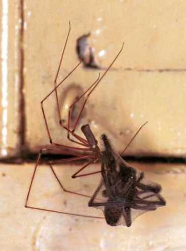
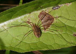
The Opiliones, or daddy long legs and harvestmen "spiders", is a large group (order) with a worldwide distribution.
The difference between the daddy-longlegs and the harvestman is that in harvestman the cephalothorax and the abdomen is almost fused together which looks like one structure whereas in daddy long leg spiders the cephalothorax and the abdomen are two distinct features connected by a visible narrow tube.
Daddy long leg spiders are harmless to man as their jaws are unable to penetrate human skin and the venom dose, although a neurotoxin is also too minute. Normally the spider throws tough stiff web material over the victim and disables its mobility. After the prey is motionless, the spider engulfs its prey by spinning the web all around it. The spider makes a hole in the ball of the web and bites with its small jaws in the weaker parts of the prey. Then it spews digesting juices in the wound.
When ready to copulate, the male spider makes couple of silk threads, then places his genital area on the thread and starts rubbing along the thread which stimulate his sexual organs. This action produces secretion containing sperm, which is drawn toward his poison fangs and is sucked in by his palps. When he finds the female, he vibrates his whole body on her web. As the female approaches, the male strokes her first pair of legs. He then inserts both of his palps in the female vulva.
Harvestmen have an oval shaped body. The front and back of the body is grown together in contrast to true spiders where the front and back end is separated by a stalk (pedicel). They have two eyes in the middle of their head looking sideways. Most of the harvestmen have long legs, but not all.
Many harvest men are omnivorous, feeding upon small invertebrates as well as fruits and vegetables. Pedipalps catch the food before passing it to the chelicerae where it is held and crushed. It then passes through the mouth with the help of suction from the pharynx, and the food is ingested, for the most part, inside the arachnid.
Harvestmen do not possess poison glands but has instead glands that produce a stinky odor. They also have no silk glands or spinners.
Copulation is directly with a penis.
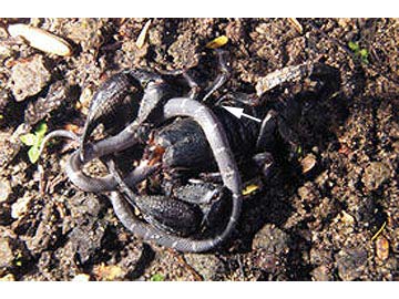
Scorpions (old order Scorpionida), eurypterids and pseudoscorpions
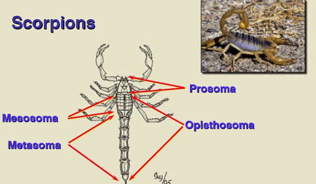
Scorpions also have two main body regions, although the abdomen is sometimes divided into more.
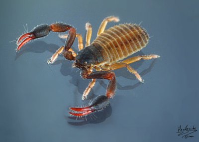
Most are aggressive feeders, capturing prey by means of large pincers.
Scorpions perform a courtship dance during which the male deposits sperm within the female's genital pore. Newly hatched scorpions are immature and ride on the mother's back until after their first molt.
The scorpion tail is equipped with a venomous stinger that is used to paralyze or kill prey that is difficult to subdue.
Scorpion venom usually causes no more injury than a bee or wasp sting, but there are species in Arizona, Mexico and North Africa that can cause death.
The oldest fossils for scorpions are about 430 million years old and had gills instead of book lungs.
There are a number of clades within the arachnida (old orders) that have a scorpion type body. A rather unknown but common scorpion relative is the pseudoscorpion (old order Pseudoscorpions). This small creature is a few millimeter long and lives between detritus like leaves, bark, moss, mole-and bird nests.
They have relatively long scissors that can be as long as the rest of the body for the males. They do not have tail and no sting.
Male pseudoscorpions deposit a spermatophore on the substrate, and the female is attracted to it by scent or in some advanced species, the female is actively maneuvered to the spermatophore by the male, who aids her it its uptake. It has been found that in fights (over females, of course) between male pseudoscorpions, the male with the largest pedipalps almost always wins. However, males with big pedipalps may be disadvantaged in having a slower rate of development and reduced abilities to capture prey and dispersephoresy, a form of hitchhiking. Typically, the pseudoscorpion waits for a large beetle or fly that has come to deposit its eggs and, at an opportune moment, grabs the leg of its "ride" and holds fast while being air-transported to a new location.
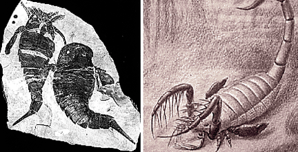
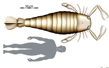
Eurypterids are an extinct group with a body plan similar to scorpions (now considered a major clade (class) by themselves or Eurypterida*). They appeared in the Ordovician and some were as long as eight feet.
*Your book has a mistake on page 403 and places this group in with the horse shoe crabs even after stating there are only 4 species in Merostomata.
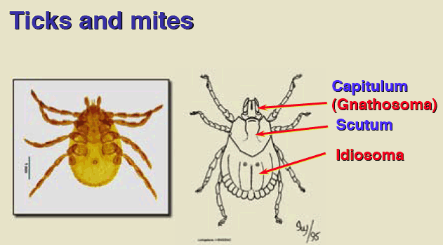
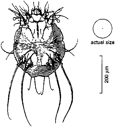
Acari (highly modified Arachnids)
With small size has come morphological change: the segmentation of the opisthosoma (abdomen) and the distinction between prosoma (cephalothorax) and opisthosoma has been almost completely lost, as have the heart and eyes in many species.
Some mites have even reduced or lost one, two, or three pairs of legs.
The oldest known mite fossil are from the Late Devonian (417 to 354 million years ago). However, because of their small size, the Acari are uncommon in the fossil record.
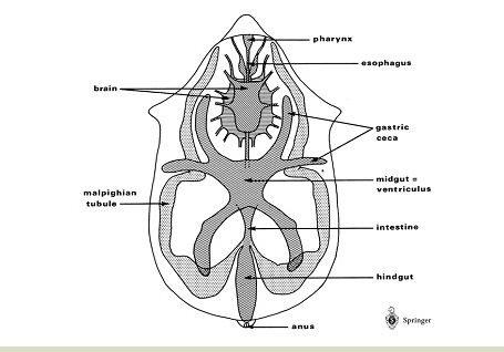
Mite internal anatomy
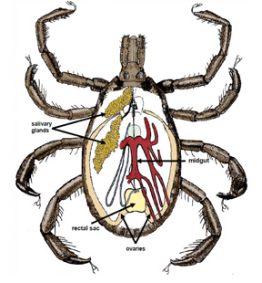
Tick internal anatomy
Mites and ticks which feed on vertebrate hair or blood often carry disease organisms, such as spirochete bacteria, responsible for relapsing fever and Lyme disease.
Others are rather unpleasant parasites themselves, such as ticks, chiggers, and the skin mites that cause mange and scabies.
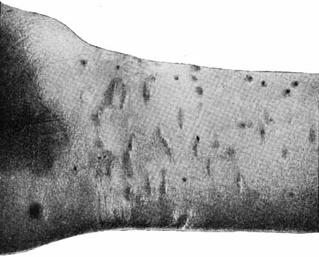
Scabies
Practically the whole life cycle is spent on and in the skin of humans. In order to feed and lay eggs, fertilized females burrow winding tunnels in the surface of the skin. The tunnels are extended by 1-5 mm a day and can be seen on the skin as very thin twisting lines a few millimeters to several centimeters long.
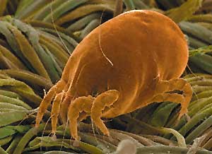
Dust mites:
In homes, the highest concentration of dust mites can be found in mattresses and upholstered furniture. A typical profile of dust mite allergen location might be 10,000 nanograms per gram (ng/g) – mattress, 8,000 ng/g – upholstered chair, 2,000 ng/g – carpet, 1,000 ng/g – draperies. As many as 2,000,000 live mites may inhabit the average mattress. With the average pillow, 30% of its weight is comprised of dead human skins scales and dust mite allergen.
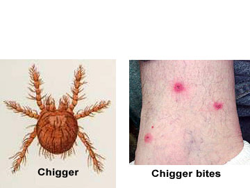
Chiggers
Chiggers are a type of mite in which the larval stage feeds on skin, causing welts and severe itching.
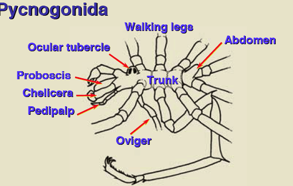
4. Sea spiders (Pycnogonida)
Body not divided into distinct regions. Unique proboscis at the anterior end. The earliest fossils are known from the Cambrian.
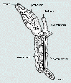
Sea spiders are carnivorous, some feeding on other invertebrates by sucking out the juices, while others tear their prey apart and pass it into a proboscis for feeding. The digestive system extends into the legs, and the pair of simple eyes are positioned near the end of the trunk .
Most sea spiders are 1 to 10 mms in length but some deepwater species in Antarctica can have a leg span up to 90 cms!
Pycnogonids have extremely reduced bodies in which the abdomen has almost disappeared, while the legs are long and clawed. The head has a long proboscis with an unusual terminal mouth and several simple eyes on a central tubercle. The pharynx not only acts as a pump, but also macerates the food by means of bristles projecting into the lumen from the plates. Because of the lack of space in the tiny body, parts of some organ systems, such as the digestive and reproductive systems, are housed in the legs. Diverticula (digestive ceca) from the gut extend far into legs and the female legs are expanded to provide room for the developing ova
The head also bears a pair of claws and a pair of ovigers on which the eggs are carried. Pycnogonids are one of the few groups of animals in which the males exclusively care for the developing eggs The larva either remains on the ovigerous legs of the male or more frequently becomes an ectoparasite or endoparasite on hydroids or corals. In either case the larva is followed by a four-legged and then a six-legged form each with a further pair of posterior buds. When the adult form is reached some appendages may be lost.
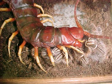
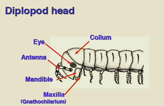
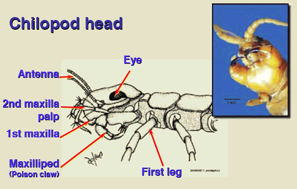
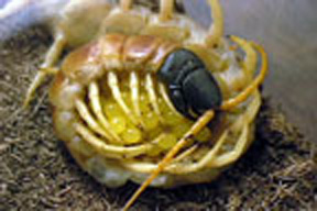
6. Myriapoda: centipedes and millipedes
The myriapods are divided into the centipedes which are active predators and the millipedes which eat plant material.
The name centipede is derived from the Latin words "centi" (100) and "pede" (foot). Centipedes have pair of poison claws behind the head, (modified first pair of thoracic legs) and use the poison to paralyze their prey, usually small insects. Like scorpions, the centipedes can subdue some surprisingly aggressive prey. Only the larger centipedes pose any danger to man.
The class is distinguished by division into two tagmata, head and a flattened multi segmented trunk. Head posses modified appendages such as mandibles and maxillae.
Centipedes mate in the manner typical of many arthropods -- the male produces a packet of sperm called a spermatophore, which is taken up by the female and stored in a pair of internal sacs known as spermathecae, where it is used to fertilize her eggs. In some species, the spermatophore is inserted directly into the female’s body by the male; in most, the spermatophore is simply left lying on the ground where it is later found and picked up by the female. Females are capable of maintaining live sperm in their spermathecae for a considerable length of time, and can still lay viable eggs over six months after their last mating.
The name millipede is derived from the Latin words "milli" (1000) and "pede" (foot). In some millipedes, the male simply deposits a spermatophore on the ground and leaves it for the female to find later. She picks up the spermatophore and inserts it into her spermathecae. In other species, the male and female pair will coil around each other so their sex organs meet, and the male will use the legs on his tenth segment to place the spermatophore inside the female.
In some species of millipedes, adult males may undergo a transition known as "periodomorphosis", in which a sexually mature male molts and emerges as an immature male with undeveloped sex organs. He later molts again and re-emerges as a sexually mature male once again. It is not known why this process takes place. It may be a response to adverse environmental conditions.
Excretion is accomplished via Malphigian tubules and trachea allow for gas exchange.
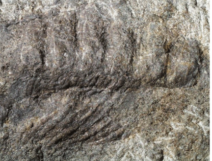
The oldest fossils are Myriapod-like marine creatures from Cambrian-age strata. Fossil burrows in Ordovician strata have led to conjecture that Myriapods might have been living on land as early as 400 million years ago. The oldest myriapod in the fossil record, and possibly the oldest known terrestrial air-breathing organism is the millipede Pneumodesmus newmani from the mid Silurian dating to about 425 million years. It exhibits cuticular openings that taxonomists have interpreted as spiracles.
 bottom dweller
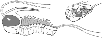 pelagic