Chordate lab
This is an observational lab devoted to the rest of the deuterostome line.
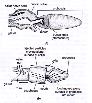
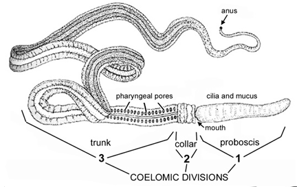
Hemichordates are distinguished by a tripartite (threefold) division of the body. At the forward end of the body is a preoral lobe, behind this is a collar, and last comes a trunk (so 3 segments). Each contains a division of the coelom. The name "hemichordate" means "half chordate," and hemichordates share some (but not all) of the typical chordate characteristics. There are branchial openings, or "gill slits," that open into the pharynx; there is a rudimentary structure in the collar region, the stomochord, that is considered similar to a notochord by some authors (although develops differently from a notochord in the embryo and involves different genes); and there is a dorsal nerve cord, in addition to a smaller ventral nerve cord.
However, hemichordates are not classified as true chordates, although they are quite closely related. Some DNA-based studies of evolution suggest that hemichordates are actually closer to echinoderms than to true chordates. This is supported by the fact that the larvae of at least some hemichordates look very much like those of some echinoderms.
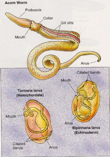
Acorn worms: We usually have acorn worms in the laboratory for students to view.
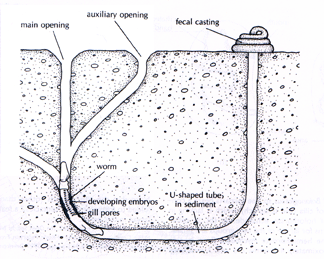
Acorn worms live on the sea-bed, from the shoreline down to the depths of 10,000 ft.. (3,050 m)The skin is covered with cilia as well as glands that secrete mucus. Some produce a bromide compound that gives them a medicinal smell and might protect them from bacteria and predators.
Acorn worms move by cilia movements and body contractions. The stomochord provides support for the front of the animal and the coelom acts as a hydrostatic skeleton for the back. They are filter feeders.
Acornworms do not have rich fossil records, though fossils have been recognized from the Lower Triassic some 250 million years ago
Chordata
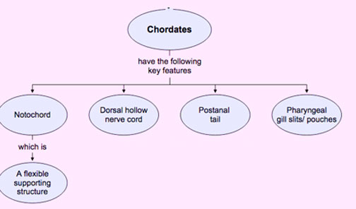
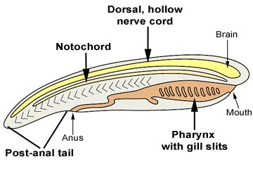
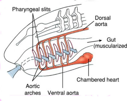
All members of clade chordata have four main characteristics that distinguish them from other animals. Although many chordate animals lack one or more of these characteristics as adults, all animals exhibit them at some stage of their life cycle.
The defining chordate characteristic is the notochord, a fairly stiff rod of cartilage (http://en.wikipedia.org/wiki/Cartilage) that runs along the dorsal side of the body. The notochord is formed during development of the embryo, from the roof of the embryonic gut. It serves to stiffen the body, thus facilitating an efficient, swimming motion.
The nerve cord is located just above the notochord. This dorsal location differs from that of other animals, such as the annelids and arthropods. In most chordate animals, the anterior end of the nerve cord is expanded to form a brain.
Pharyngeal gill slits allow water entering the mouth to exit from the pharynx, rather than through the anus. This water flow is used for filter-feeding and/or respiration in the invertebrate chordates (homologous to gills in fish).
The post-anal tail is composed mainly of muscle and provides additional propulsion for swimming.
Urochordata =Tunicata
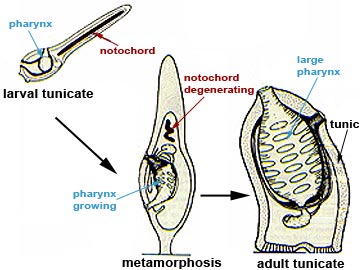
We used to think of the tunicates as a rather "primitive" form of chordate .Their specific larva that looks a lot like a small, simplified fish. They probably would be better considered as a successful lineage all their own, derived from a stock that gave rise, in time, to actual chordates, but not through the tunicates.
All tunicates are marine animals and while none is found in freshwater, a relatively large number of species may live in estuaries. There are three subgroups of tunicates possessing some common characteristics.
Larvae
As do many other invertebrate groups, they possess a larva that undergoes a dramatic metamorphosis into a juvenile. Their larvae look like small tadpoles or very simplified small fishes. Having such a notochord in the tail allows them to swim much more efficiently than they could without it. The larvae and the swimming ability are transitory. Although the gut, branchial basket, and gill slits are present, they are not functional and do not open in the larva. Only in one class, the Larvacea, do the adults retain the tadpole shape, and even so, as adults they do not swim much.
Adults
Showing other similarities to the chordates, all tunicates possess openings in the front part of their gut that are considered to be homologous with the gill slits of simple vertebrates such as lampreys. Additionally, most adult tunicates possess a rudimentary nervous system; as with many of their structures, however, the larval nervous system is more sophisticated, consisting of a main dorsal hollow nerve cord located above the notochord with very small nerves leaving it. This is a pattern also seen in the chordates.
As their name implies, many possess a "tunic" or outer supportive/protective layer. This is comprised of a type of cellulose called tunicin. Curiously, one of the few other places where cellulose is found in animals is as fibers deposited within the skin.
Only the two siphonal openings, an incurrent and an excurrent for water passage into and out of the branchial basket, pierce the tunic. Unlike other animals with a surrounding cuticle, tunicates grow inside the tunic without molting. This is probably due to their ability to resorb and redeposit tunic materials at the mantle-tunic interface. Some species have channels in the tunic that are continuous with internal blood spaces, and allow for the secretion and redeposition of tunic material as well as for the secretion of defensive materials into the tunic.
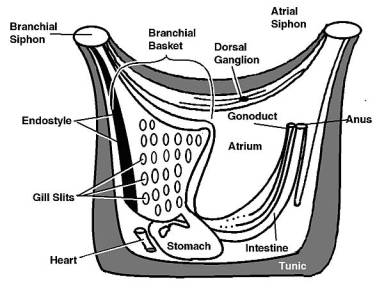
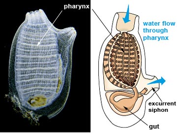
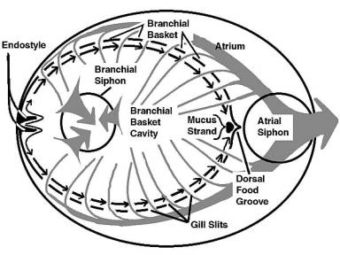
Cilia lining the edges of the gill slits pump water through the branchial basket. In this structure, mucus is produced from a ventral groove or gutter called the endostyle. A primary component of this mucus is indistinguishable from vertebrate thyroid hormone, and the endostyle is often regarded as the thyroid gland's evolutionary precursor. The mucus flows as a sheet from the ventral groove dorsally over the branchial basket's inner surfaces. The mucus, with its adherent food, is moved by ciliary action to a food groove located on the dorsal midline of the branchial basket.
From there it is moved, in the food groove, into the mouth. A very short esophagus transfers the food-laden mucus to a bag-like stomach. Associated with the stomach is a large structure called the digestive gland. The function of the digestive gland is not fully understood, but it probably contributes to digestion and absorption of food. The short, straight intestine leaves the stomach and leads to a rectum and anus. Feces are deposited in the atrium, and flushed out with the excurrent water passing out the excurrent siphon.
Tunicates are effective suspension-feeders; even small ones can filter hundreds of liters of water per day and remove well over 95% of its bacteria.
We have sea squirts in the laboratory.
The tunicates possess an open circulatory system; one that is largely without vessels. Blood flows through large tissue spaces or blood channels in the tissues. Arteries, veins, and capillaries are usually absent. They have a heart, but it is a simple tube with walls that contract to force the blood through it. Unlike the heart in most animals, this heart is capable of reversing its beat. Generally, the heart beats about a hundred times in one direction, stops for a moment and then beats about a hundred times in the other direction.
In the film below which shows the heart from two different angles, play close attention aroung 2.04 minutes where the heart stops and starts beating in the other direction.
In some squirts, the blood contains odd rare-earth chemicals, commonly vanadium or niobium. These metals were once thought to assist in respiration, but are now known to be anti-fouling or anti-predator defenses . Other chemicals that appear to be primarily defensive are found in the blood. Blood serum containing these metals and other noxious chemicals either leaks through the epidermis, or is secreted by it and oozes through the tunic. Additional, exceedingly acidic, defensive chemicals are found in the tunic secretions of a few species
All tunicates are hermaphroditic, and are often self-fertile. The gonads can develop just about anywhere in the animal. The gonoducts run parallel to the intestine and empty into the atrium near the anus. Gametes released from the gonoducts develop into non-feeding "tadpole" larvae that eventually locate a place to settle and transform into adults.
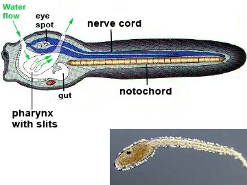
Clade (Phylum) Chordata: subphylum Cephalochordata
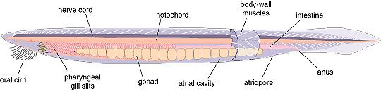
The cephalochordates have all the typical chordate features. The dorsal nerve cord is supported by a muscularized rod, or notochord. The pharynx is perforated by over 100 pharyngeal slits or "gill slits", which are used to strain food particles out of the water. The musculature of the body is divided up into V-shaped blocks, or myomeres, and there is a post-anal tail. All of these features are shared with vertebrates. On the other hand, cephalochordates lack features found in most or all true vertebrates: the brain is very small and poorly developed. Sense organs are also poorly developed, and there are no true vertebrae.
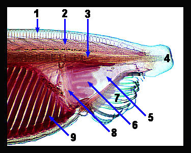
----------------------------------------------------------1. Dorsal fin and fin ray. 2. Dorsal nerve cord. 3. Notochord. 4. Rostrum.
----------------------------------------------------------5. Vestibule. 6. Wheel organ. 7. Oral cirri. 8. Velum. 9. Gill bar
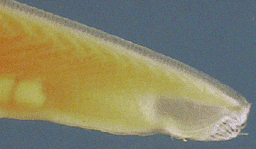
Water is taken in through the mouth, drawn in by the beating of cilia located on the wheel organ, a set of ridges lying inside the mouth. The water is first filtered by the oral cirri, slender projections that surround the opening of the mouth, clearly visible on the photograph above. It then passes through the gill slits. These gill slits are enclosed by folds of the body wall, the metapleural folds, to form a body cavity known as the atrium. Food particles in the water are trapped by mucus, while water passes through the slits and out of the atrium through the atriopore, located towards the posterior end.
The rest of the digestive system is fairly simple: a pouch or hepatic caecum secretes digestive enzymes, and actual digestion takes place in a specialized part of the intestine known as the iliocolonic ring. Cephalochordates also have a well-developed circulatory system and a simple excretory system composed of paired nephridia. The sexes are separate, and both males and females have multiple paired gonads. Eggs are fertilized externally, and develop into free-swimming, fishlike larvae.